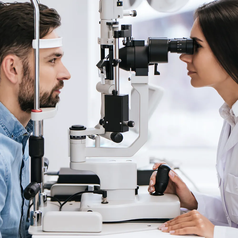
state-of-the-art technology
Dark Adaptation Testing
The AdaptDx® is the first instrument to provide a practical and objective measurement of dark adaptation function, which is severely impaired in age-related macular degeneration (AMD), retinitis pigmentosa (RP) and other macular degenerations.
The measurement of dark adaptation function provides valuable, early insight into AMD, RP and other macular degenerations. The AdaptDx is a significant advance in the measurement of dark adaptation function—the recovery of vision when going from daylight to darkness. Dark adaptation function is dramatically impaired from the earliest stages of AMD, RP and other macular degenerations, with increasing impairment as the diseases progress.
The AdaptDx is a valuable complement to existing imaging devices used in routine eye exams for macular disease assessment. It provides functional information that adds to the structural information obtained from fundus photography and OCT imaging. This additional information is particularly valuable for early assessment, as biological function is often affected earlier than structural changes can be identified.
Fundus photography
Fundus photography involves capturing a photograph of the back of the eye i.e. fundus. Specialized fundus cameras that consist of an intricate microscope attached to a flash enabled camera are used in fundus photography. The main structures that can be visualized on a fundus photo are the central and peripheral retina, optic disc and macula.
Optometrists use fundus photography for monitoring the progression of certain eye condition/diseases. Fundus photographs are also used to document abnormalities of disease process affecting the eye, and/or to follow up on the progress of the eye condition/disease such as diabetes, age-macular degeneration (AMD), glaucoma, Neoplasm of the choroid, cranial nerves, retinal or eyeball, etc.
Optical Coherence Tomography
Optical Coherence Tomography (OCT) is a non-invasive imaging test. OCT uses light waves to take cross-section pictures of your retina.
This allows your ophthalmologist to map and measure their thickness. These measurements help with diagnosis. They also provide treatment guidance for glaucoma and diseases of the retina. These retinal diseases include age-related macular degeneration (AMD) and diabetic eye disease.
OCT is often used to evaluate disorders of the optic nerve as well. The OCT exam helps your ophthalmologist see changes to the fibers of the optic nerve. For example, it can detect changes caused by glaucoma.
Order Contacts Online!
Order contacts when it's convenient for you. New and current patients can now order online anytime.
Patient Testimonials
Powered by EyeCare Specialties of Ohio
Each practice in the EyeCare Specialties network is focused on the eyes of Ohio. Our family of practices have been caring for Ohio's communities for decades. Learn More.